
Neuroimaging projects
Magnetic Resonance Imaging (MRI)
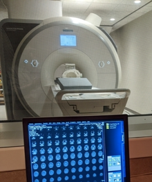
IU's state-of-the-art Imaging Research Facility (IRF) is located down the street in the Psychology building.
The IRF houses a Siemens 3 Tesla Prisma MRI scanner. Its capabilities include structural MRI, functional MRI (fMRI), diffusion and perfusion imaging, and MR spectroscopy. The lab has the capability for multisensory stimulus delivery and behavioral response collection.
Some of our projects involve the IRF, where we collect resting state functional connectivity data, diffusion imaging, and task-based fMRI. We also perform anatomical brain scans for more precise targeting in TMS studies.
Folco K, Cheng H, Newman S, Pilacinski A, Wali M, Babu R, Block HJ. (2023). Resting state functional connectivity changes among multisensory and motor regions associated with visuo-proprioceptive recalibration. Abstract for poster presentation, Society for Neuroscience Annual Meeting.
The brain estimates hand position using visual and proprioceptive (position sense) cues. When a spatial mismatch is introduced between visual and proprioceptive cues, e.g. by shifting a visual cursor away from the unseen hand, the brain recalibrates visual and proprioceptive estimates of hand position to compensate. Visuo-proprioceptive recalibration occurs in the context of visuomotor adaptation paradigms such as cursor rotation, but it is a distinct process. Visuomotor adaptation is known to involve activity in the cerebellum and primary motor cortex (M1), but little is known about the neural basis of visuo-proprioceptive recalibration itself. Under a traditional framework, M1, ventral premotor cortex (PMv), and cerebellum (CB) would be considered motor structures while primary somatosensory cortex (S1) and anterior superior parietal lobule (aSPL) would be considered sensory, but today, most would agree that sensory and motor functions emerge from distributed processing among these and other regions. M1 and S1 have both been linked to proprioceptive activity, and aSPL, PMv, and CB have been linked to multisensory (visual and proprioceptive) activity. Here we ask whether resting state functional connectivity among these regions undergoes any change following a task involving visuo-proprioceptive recalibration, but not motor adaptation. Participants completed a resting state scan, a hand position estimation task using veridical cues (baseline task), a second resting state scan, a hand position estimation task where the visual cue gradually shifted 70 mm away from the proprioceptive cue (recalibration task), and finally a third resting state scan. Using a seed-based network analysis, preliminary data (N=6) indicates that connectivity between S1, a traditionally sensory region, and PMv, a traditionally motor region, may be altered after the recalibration task relative to baseline. In addition, we found that connectivity between CB and aSPL may be related to the magnitude of proprioceptive recalibration measured behaviorally. In addition, visuo-proprioceptive recalibration in a pre-defined sensorimotor network consisting of the right anterior superior parietal lobule, M1, S1PMv, and left cerebellum lobule VI was related to changes in network measures. Taken together, these results support the idea that visuo-proprioceptive recalibration is associated with changes in resting state functional connectivity among both motor and multisensory regions.
The brain estimates hand position using visual and proprioceptive (position sense) cues. When a spatial mismatch is introduced between visual and proprioceptive cues, e.g. by shifting a visual cursor away from the unseen hand, the brain recalibrates visual and proprioceptive estimates of hand position to compensate. Visuo-proprioceptive recalibration occurs in the context of visuomotor adaptation paradigms such as cursor rotation, but it is a distinct process. Visuomotor adaptation is known to involve activity in the cerebellum and primary motor cortex (M1), but little is known about the neural basis of visuo-proprioceptive recalibration itself. Under a traditional framework, M1, ventral premotor cortex (PMv), and cerebellum (CB) would be considered motor structures while primary somatosensory cortex (S1) and anterior superior parietal lobule (aSPL) would be considered sensory, but today, most would agree that sensory and motor functions emerge from distributed processing among these and other regions. M1 and S1 have both been linked to proprioceptive activity, and aSPL, PMv, and CB have been linked to multisensory (visual and proprioceptive) activity. Here we ask whether resting state functional connectivity among these regions undergoes any change following a task involving visuo-proprioceptive recalibration, but not motor adaptation. Participants completed a resting state scan, a hand position estimation task using veridical cues (baseline task), a second resting state scan, a hand position estimation task where the visual cue gradually shifted 70 mm away from the proprioceptive cue (recalibration task), and finally a third resting state scan. Using a seed-based network analysis, preliminary data (N=6) indicates that connectivity between S1, a traditionally sensory region, and PMv, a traditionally motor region, may be altered after the recalibration task relative to baseline. In addition, we found that connectivity between CB and aSPL may be related to the magnitude of proprioceptive recalibration measured behaviorally. In addition, visuo-proprioceptive recalibration in a pre-defined sensorimotor network consisting of the right anterior superior parietal lobule, M1, S1PMv, and left cerebellum lobule VI was related to changes in network measures. Taken together, these results support the idea that visuo-proprioceptive recalibration is associated with changes in resting state functional connectivity among both motor and multisensory regions.