TMS projects

Transcranial Magnetic Stimulation (TMS)
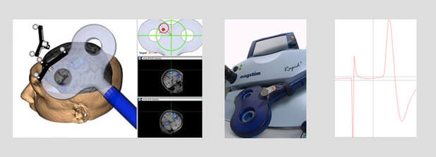
TMS is a way to non-invasively stimulate the brain. We use single and paired-pulse TMS to investigate changes in motor cortical activity in response to behavioral and neurophysiological interventions. This technique can be used to assess intracortical inhibition and neuroplasticity.
TMS can also be used repetitively (rTMS) to cause a slight temporary change in activity in a region of the cortex. We use a pattern of rTMS called theta burst stimulation (TBS) that takes 40 seconds to apply, but has effects that last up to an hour. This lets us infer whether the brain region we stimulated plays a role in the behavior being studied.
Example publications
Mirdamadi, J.L., Block, H.J. (2021). Somatosensory versus cerebellar contributions to proprioceptive changes associated with motor skill learning: A theta burst stimulation study. Cortex 140:98-109. [PubMed]
Background: It is well established that proprioception (position sense) is important for motor control, yet its role in motor learning and associated plasticity is not well understood. We previously demonstrated that motor skill learning is associated with enhanced proprioception and changes in sensorimotor neurophysiology. However, the neural substrates mediating these effects are unclear.
Objective: To determine whether suppressing activity in the cerebellum and somatosensory cortex (S1) affects proprioceptive changes associated with motor skill learning.
Methods: 54 healthy young adults practiced a skill involving visually-guided 2D reaching movements through an irregular-shaped track using a robotic manipulandum with their right hand. Proprioception was measured using a passive two-alternative choice task before and after motor practice. Continuous theta burst stimulation (cTBS) was delivered over S1 or the cerebellum (CB) at the end of training for two consecutive days. We compared group differences (S1, CB, Sham) in proprioception and motor skill, quantified by a speed-accuracy function, measured on a third consecutive day (retention).
Results: As shown previously, the Sham group demonstrated enhanced proprioceptive sensitivity after training and at retention. The S1 group had impaired proprioceptive function at retention through online changes during practice, whereas the CB group demonstrated offline decrements in proprioceptive function. All groups demonstrated motor skill learning. However, the magnitude of learning differed between the CB and Sham groups, consistent with a role for the cerebellum in motor learning.
Conclusion: Overall, these findings suggest that the cerebellum and S1 are important for distinct aspects of proprioceptive changes during skill learning.
Mirdamadi, J.L., Seigel, C.R., Husch, S.D., Block, H.J. (2022). Somatotopic specificity of neural and perceptual changes in spatial estimation of the hand. Cerebral Cortex 32(6): 1184-1199. [PubMed]
Abstract: When visual and proprioceptive estimates of hand position disagree (e.g., viewing the hand underwater), the brain realigns them to reduce mismatch. This perceptual change is reflected in primary motor cortex (M1) excitability, suggesting potential relevance for hand movement. Here, we asked whether fingertip visuo-proprioceptive misalignment affects only the brain's representation of that finger (somatotopically focal), or extends to other parts of the limb that would be needed to move the misaligned finger (somatotopically broad). In Experiments 1 and 2, before and after misaligned or veridical visuo-proprioceptive training at the index finger, we used transcranial magnetic stimulation to assess M1 representation of five hand and arm muscles. The index finger representation showed an association between M1 excitability and visuo-proprioceptive realignment, as did the pinkie finger representation to a lesser extent. Forearm flexors, forearm extensors, and biceps did not show any such relationship. In Experiment 3, participants indicated their proprioceptive estimate of the fingertip, knuckle, wrist, and elbow, before and after misalignment at the fingertip. Proprioceptive realignment at the knuckle, but not the wrist or elbow, was correlated with realignment at the fingertip. These results suggest the effects of visuo-proprioceptive mismatch are somatotopically focal in both sensory and motor domains.
Abstract: When visual and proprioceptive estimates of hand position disagree (e.g., viewing the hand underwater), the brain realigns them to reduce mismatch. This perceptual change is reflected in primary motor cortex (M1) excitability, suggesting potential relevance for hand movement. Here, we asked whether fingertip visuo-proprioceptive misalignment affects only the brain's representation of that finger (somatotopically focal), or extends to other parts of the limb that would be needed to move the misaligned finger (somatotopically broad). In Experiments 1 and 2, before and after misaligned or veridical visuo-proprioceptive training at the index finger, we used transcranial magnetic stimulation to assess M1 representation of five hand and arm muscles. The index finger representation showed an association between M1 excitability and visuo-proprioceptive realignment, as did the pinkie finger representation to a lesser extent. Forearm flexors, forearm extensors, and biceps did not show any such relationship. In Experiment 3, participants indicated their proprioceptive estimate of the fingertip, knuckle, wrist, and elbow, before and after misalignment at the fingertip. Proprioceptive realignment at the knuckle, but not the wrist or elbow, was correlated with realignment at the fingertip. These results suggest the effects of visuo-proprioceptive mismatch are somatotopically focal in both sensory and motor domains.
